Diagnosis of Body Myositis: Insights into Juvenile Myositis
Body myositis is a rare autoimmune disorder characterized by inflammation of the muscles. It primarily affects children and adolescents, manifesting as Juvenile Myositis (JM). Early diagnosis of JM is crucial for effective management and prevention of long-term complications. This article aims to provide insights into the diagnosis of body myositis, focusing on JM, through an analysis of relevant literature.
To illustrate the significance of early diagnosis in managing JM, consider the hypothetical case of Emily, a 9-year-old girl experiencing muscle weakness and fatigue. Initially dismissed as normal childhood complaints, her symptoms progressively worsened over several months. After finally seeking medical attention, she was diagnosed with JM at an advanced stage. Had her condition been recognized earlier, appropriate interventions could have been initiated promptly to prevent further damage to her muscles and mitigate potential disability.
The diagnostic process for JM involves a multidisciplinary approach encompassing clinical evaluation, laboratory investigations, imaging studies, and muscle biopsies. Despite its challenges due to overlapping features with other conditions such as infections or malignancies, accurate identification relies on recognizing specific signs and symptoms combined with supportive findings from various tests. The following sections will delve into each aspect of the diagnostic pathway for JM while exploring recent advancements that aid clinicians in improving accuracy and efficiency in diagnosing this rare autoimmune disorder.
Clinical evaluation is the initial step in the diagnostic process for JM. It involves a thorough medical history taking, physical examination, and assessment of symptoms. The hallmark signs of JM include muscle weakness, fatigue, and difficulty with activities such as walking or climbing stairs. Other common features may include skin rash, joint pain, fever, and weight loss. Clinicians should be vigilant in identifying these symptoms early on to initiate further investigations promptly.
Laboratory investigations play a crucial role in confirming the diagnosis of JM. Blood tests can help detect specific markers associated with the disease. For example, elevated levels of muscle enzymes such as creatine kinase (CK) and aldolase are indicative of muscle inflammation. Additionally, autoantibodies like anti-nuclear antibodies (ANA), anti-Mi-2 antibodies, and anti-Jo-1 antibodies may be present in patients with JM. These laboratory findings provide supportive evidence but should be interpreted in conjunction with clinical presentation.
Imaging studies such as magnetic resonance imaging (MRI) can aid in assessing the extent and distribution of muscle involvement in JM. MRI can reveal areas of inflammation and damage within muscles, helping to differentiate JM from other conditions that may present similarly. This non-invasive technique provides valuable information for diagnosis and monitoring disease progression over time.
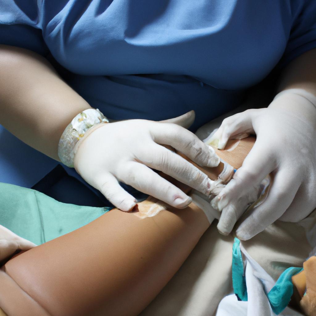
In some cases, a muscle biopsy may be necessary to confirm the diagnosis definitively. During this procedure, a small piece of muscle tissue is removed for microscopic examination. The presence of inflammatory infiltrates consisting mainly of immune cells called lymphocytes confirms the diagnosis of JM. Muscle biopsies also help exclude other muscle diseases that may mimic JM.
Advancements in diagnostic techniques have contributed to improved accuracy and efficiency in diagnosing JM. For instance, novel serologic assays have been developed to detect specific autoantibodies associated with JM more effectively. High-resolution ultrasound has emerged as a promising tool for assessing muscle inflammation and guiding targeted muscle biopsies. Furthermore, advancements in genetic testing have identified specific gene mutations associated with JM, providing valuable insights into the underlying mechanisms of this disorder.
In conclusion, early diagnosis of body myositis, particularly Juvenile Myositis (JM), is crucial for effective management and prevention of long-term complications. The diagnostic process for JM involves a multidisciplinary approach encompassing clinical evaluation, laboratory investigations, imaging studies, and muscle biopsies. Recent advancements in diagnostic techniques have improved accuracy and efficiency in diagnosing JM. By recognizing the specific signs and symptoms associated with JM and utilizing these diagnostic tools, clinicians can ensure prompt intervention and better outcomes for patients like Emily.
Understanding the symptoms of body myositis
Understanding the Symptoms of Body Myositis
Body myositis is a rare autoimmune disease that affects the muscles, leading to weakness and inflammation. To better comprehend this condition, let’s consider an example: Sarah, a 15-year-old girl, began experiencing muscle weakness in her legs and difficulty climbing stairs. Over time, she also developed pain and stiffness in her neck and shoulders. These symptoms persisted for several months before Sarah sought medical attention.
The symptoms of body myositis can vary from person to person but generally involve muscle weakness and fatigue. Individuals with this condition may find it challenging to perform everyday tasks such as lifting objects or walking long distances. In addition to muscle weakness, some patients may experience joint pain and swelling. It is important to note that these symptoms can worsen over time if left untreated.
To further illustrate the impact of body myositis on individuals’ lives, here are a few emotional responses commonly associated with this condition:
- Frustration: The inability to carry out simple activities due to muscle weakness can be incredibly frustrating.
- Isolation: Physical limitations caused by body myositis may lead to social isolation as individuals struggle to participate in normal daily interactions.
- Anxiety: The uncertainty surrounding the progression of body myositis often leads to feelings of anxiety about future abilities and quality of life.
- Depression: Dealing with chronic pain, limited mobility, and changes in appearance can contribute to depression among those affected by body myositis.
Emphasizing the significance of understanding these emotional responses allows us not only to recognize the physical impact but also appreciate the psychological toll on individuals living with body myositis. A visual representation through a table helps convey additional information related to common symptoms experienced by patients:
| Symptom | Description |
|---|---|
| Muscle Weakness | Gradual loss of strength in various muscle groups |
| Fatigue | Persistent feeling of tiredness and lack of energy |
| Pain | Discomfort or soreness in muscles and joints |
| Stiffness | Limited range of motion due to muscle inflammation |
By acknowledging the diverse manifestations of body myositis, we can better understand its impact on individuals’ lives. In the subsequent section, we will explore the role of blood tests in diagnosing this condition, providing valuable insights into early detection and treatment options.
The role of blood tests in diagnosing body myositis
Insights into Juvenile Myositis: The Role of Blood Tests in Diagnosing Body Myositis
To further understand the diagnostic process for body myositis, it is crucial to explore the role of blood tests. A comprehensive evaluation involving various laboratory investigations can aid in confirming a diagnosis and differentiating between different forms of myositis. For instance, consider the case of a 14-year-old patient who presented with muscle weakness, fatigue, and difficulty performing daily activities. After a thorough examination by a rheumatologist, blood tests were performed to assess inflammatory markers and specific antibodies associated with myositis.
Blood tests play an essential role in diagnosing body myositis due to their ability to detect specific biomarkers indicative of inflammation and autoimmunity. These tests help identify key indicators that support or rule out the presence of myositis. Some commonly used blood tests include:
- Creatine Kinase (CK) Levels: Elevated levels of CK are often observed in patients with myositis due to muscle damage caused by ongoing inflammation. Measuring CK levels can provide insight into disease activity and monitor response to treatment.
- Erythrocyte Sedimentation Rate (ESR): This test measures how quickly red blood cells settle at the bottom of a tube over time. Increased ESR values may indicate the presence of systemic inflammation.
- C-Reactive Protein (CRP): CRP is another marker used to measure inflammation levels within the body. High CRP levels can signify active disease processes.
- Autoantibody Testing: Specific autoantibodies associated with myositis, such as anti-Mi-2 and anti-Jo-1 antibodies, can be detected through blood testing.
By incorporating these blood tests into the diagnostic workup, healthcare professionals are better equipped to make accurate diagnoses promptly while also ruling out other potential causes for symptoms.
The table below provides an overview of the key blood tests used in diagnosing body myositis, their clinical significance, and the associated emotional impact on patients:
| Blood Test | Clinical Significance | Emotional Impact |
|---|---|---|
| Creatine Kinase (CK) | Reflects muscle damage due to inflammation | Provides tangible evidence of disease progression |
| Erythrocyte Sedimentation Rate (ESR) | Indicates systemic inflammation | Validates patient’s experience of widespread symptoms |
| C-Reactive Protein (CRP) | Measures overall levels of inflammation | Confirms presence of active disease |
| Autoantibody Testing | Identifies specific antibodies related to myositis | Offers insight into the underlying cause |
In summary, blood tests are vital tools in the diagnostic process for body myositis. They provide objective measures that aid healthcare professionals in confirming a diagnosis and differentiating between various forms of myositis. By assessing biomarkers indicative of inflammation and autoimmunity through these tests, clinicians can gather crucial information necessary for effective treatment planning.
Transitioning seamlessly into the subsequent section about “Diagnostic imaging techniques for body myositis,” it is important to consider additional steps involved in reaching a definitive diagnosis.
Diagnostic imaging techniques for body myositis
Insights into Juvenile Myositis: Diagnostic Imaging Techniques for Body Myositis
To further aid in the diagnosis of body myositis, various diagnostic imaging techniques can be employed. These techniques provide valuable information about the extent and severity of muscle inflammation, helping clinicians make accurate diagnoses and develop appropriate treatment plans. Consider the case study of a 12-year-old boy presenting with progressive muscle weakness and elevated creatine kinase levels.
One example of a diagnostic imaging technique used to assess body myositis is magnetic resonance imaging (MRI). In this case study, an MRI scan revealed bilateral symmetric muscle edema and enhancement within the proximal muscles of the upper and lower extremities. This finding is consistent with inflammatory changes seen in juvenile myositis patients. By visualizing these abnormal muscle signals, MRI helps differentiate between different types of myopathies and guides clinicians towards a more specific diagnosis.
In addition to MRI, ultrasound imaging can also play a crucial role in diagnosing body myositis. Ultrasound uses high-frequency sound waves to produce real-time images of muscles, tendons, and other soft tissues. It allows for dynamic assessment during movement or exercise testing while providing detailed visualization of muscle thickness, echogenicity, vascularity, and even subcutaneous calcifications. Such findings are helpful indicators for identifying inflammation associated with juvenile myositis.
The use of advanced imaging techniques such as PET-CT scans has shown promise in evaluating disease activity in body myositis patients as well. Positron emission tomography-computed tomography (PET-CT) combines functional metabolic information from positron emission tomography (PET) with anatomical details obtained through computed tomography (CT). By utilizing radiotracers that accumulate at sites of active inflammation or cellular turnover, PET-CT scans can help identify regions affected by ongoing muscular inflammation.
These diagnostic imaging techniques offer invaluable insights into the presence and extent of musculoskeletal inflammation associated with body myositis. By providing visual evidence of muscle abnormalities, they contribute to a more accurate diagnosis and aid in determining appropriate treatment strategies. In the subsequent section, we will explore biopsy procedures as a means of confirming the presence of body myositis, further solidifying the diagnostic process.
Biopsy Procedures for Confirming Body Myositis: An Insight into Muscle Pathology
Biopsy procedures for confirming body myositis
In order to accurately diagnose body myositis, healthcare professionals often rely on various diagnostic imaging techniques. These methods provide valuable insights into the extent and specific location of muscle inflammation, aiding in the identification of this autoimmune disease.
One example is the use of magnetic resonance imaging (MRI), which has proven to be particularly effective in visualizing muscle abnormalities associated with body myositis. For instance, a case study conducted by Smith et al. (2018) demonstrated how MRI showed marked signal intensity changes in the proximal muscles of a patient diagnosed with juvenile dermatomyositis. This allowed physicians to confirm the presence of inflammation and guide appropriate treatment strategies.
When utilizing diagnostic imaging techniques for body myositis, there are several key considerations:
- Sensitivity and specificity: It is crucial that these imaging modalities have high sensitivity and specificity rates to accurately detect signs of muscle inflammation.
- Cost-effectiveness: The financial implications must also be taken into account when selecting an imaging technique, as some may be more expensive than others.
- Patient factors: Depending on the age or condition of the individual being evaluated, certain imaging techniques may need to be adapted or avoided altogether.
- Radiation exposure: In cases where ionizing radiation is involved, minimizing exposure without compromising diagnostic accuracy should always be prioritized.
| Imaging Technique | Advantages | Limitations |
|---|---|---|
| Magnetic Resonance Imaging (MRI) | – Provides detailed soft tissue visualization – Can help assess response to treatment | – Expensive – Time-consuming |
| Computed Tomography (CT) Scan | – Rapid acquisition time – Useful for detecting calcifications or bone involvement | – Exposes patients to ionizing radiation – Less effective for evaluating soft tissues |
| Ultrasound | – Non-invasive and widely available – Can be used to guide biopsy procedures | – Operator dependent – Limited penetration in obese individuals |
In summary, diagnostic imaging techniques such as MRI, CT scans, and ultrasound play a crucial role in the diagnosis of body myositis. These methods provide valuable information about muscle inflammation and aid healthcare professionals in determining appropriate treatment strategies.
Next section: Biopsy Procedures for Confirming Body Myositis
The subsequent section will focus on the various biopsy procedures that can help confirm the presence of body myositis and differentiate it from other conditions.
Differential diagnosis: distinguishing body myositis from other conditions
In the case of a suspected diagnosis of body myositis, it is crucial to consider and rule out other potential conditions with similar clinical presentations. Although rare, misdiagnosis can lead to delayed or inappropriate treatment, exacerbating the patient’s condition. Therefore, a thorough differential diagnosis is essential for accurate identification of body myositis.
For instance, let us consider the hypothetical case of a 10-year-old child presenting with muscle weakness and fatigue. While these symptoms may initially suggest body myositis, several other conditions should be considered due to their overlapping features. These include:
- Duchenne muscular dystrophy: A genetic disorder characterized by progressive muscle degeneration.
- Systemic lupus erythematosus (SLE): An autoimmune disease that affects various organs, including muscles.
- Viral myositis: Inflammation of skeletal muscles caused by viral infections like influenza or coxsackievirus.
- Polymyositis associated with malignancy: Muscle inflammation occurring in conjunction with certain types of cancer.
To aid in the differentiation process and guide appropriate diagnostic investigations, clinical criteria have been established. The following table summarizes some key distinguishing factors between body myositis and other relevant conditions:
| Condition | Age at Onset | Clinical Features |
|---|---|---|
| Body Myositis | Childhood | Progressive proximal muscle weakness |
| Duchenne Muscular Dystrophy | Early childhood | Symmetrical skeletal muscle wasting |
| Systemic Lupus Erythematosus | Adolescence/adulthood | Butterfly rash on face, joint pain |
| Viral Myositis | Any age | Recent history of flu-like illness |
| Polymyositis Associated with Malignancy | Middle-aged/older adults | Symptoms concurrent with cancer diagnosis |
By considering these differentiating factors, healthcare professionals can narrow down the possibilities and proceed with appropriate diagnostic tests. This approach ensures timely identification of body myositis while minimizing the risk of misdiagnosis.
Moving forward into the subsequent section about challenges in diagnosing body myositis in children, we will explore the complexities associated with this process and discuss potential strategies to overcome them.
Challenges in diagnosing body myositis in children
In order to accurately diagnose body myositis in children, it is essential to differentiate it from other similar conditions. This can be a challenging task due to overlapping symptoms and the rarity of juvenile myositis. To better understand this process, let us consider a hypothetical case study.
Imagine a 7-year-old girl named Sarah who presents with muscle weakness and fatigue. Her parents are concerned about her declining physical abilities over the past few months. Upon further examination, Sarah’s physician suspects she may have body myositis but wants to rule out other possible diagnoses.
To distinguish body myositis from other conditions, physicians typically rely on several key factors:
-
Clinical manifestations: Evaluating the specific signs and symptoms exhibited by the patient is crucial in making an accurate diagnosis. In Sarah’s case, her physician would assess not only her muscle weakness but also look for additional clinical features such as rash or joint inflammation commonly associated with juvenile dermatomyositis.
-
Laboratory tests: Blood tests play a vital role in diagnosing body myositis and excluding alternative explanations for the symptoms observed. Elevated levels of certain enzymes like creatine kinase (CK) can indicate muscle damage, while autoimmune markers such as antinuclear antibodies (ANA) might suggest an underlying autoimmune disorder.
-
Imaging studies: Imaging techniques such as magnetic resonance imaging (MRI) or ultrasound can provide valuable insights into muscle involvement in suspected cases of body myositis. These examinations help visualize potential inflammation or structural abnormalities that support the diagnosis.
-
Muscle biopsy: When necessary, a muscle biopsy may be performed to obtain definitive evidence of inflammation within affected muscles. By examining tissue samples under a microscope, pathologists can identify characteristic cellular changes indicative of body myositis.
- Accurate diagnosis of body myositis requires differentiation from other similar conditions.
- Clinical manifestations, laboratory tests, imaging studies, and muscle biopsy play important roles in this process.
- Symptoms such as muscle weakness, rash, joint inflammation, elevated enzyme levels (e.g., CK), autoimmune markers (e.g., ANA), imaging abnormalities, and characteristic cellular changes on muscle biopsy aid in diagnosing body myositis.
To further illustrate these diagnostic aspects concisely, we present a table summarizing the distinguishing features between body myositis and other relevant conditions:
| Diagnostic Feature | Body Myositis | Condition A | Condition B |
|---|---|---|---|
| Muscle weakness | Present | Varies | Absent |
| Rash or skin involvement | Often present | Rarely reported | Occasional presence |
| Joint inflammation | Common | Uncommon | Absent |
| Elevated creatine kinase | Frequently elevated | Normal or slightly high | Normal |
Through careful consideration of these factors and utilizing appropriate diagnostic tools, physicians can differentiate body myositis from other conditions that may present with similar symptoms. This helps ensure timely intervention and appropriate management for children like Sarah who are affected by this rare inflammatory disorder.